Overview
Our minimal access gynaecology service uses a range of medical and surgical options to help manage common and potentially debilitating gynaecological conditions such as fibroids, endometriosis, ovarian cysts and polyps. These conditions can have a significant impact on fertility as well as the outcome of fertility treatments such as IVF. Almost all operations are performed as ‘keyhole’ surgery (small incisions in the abdomen for laparoscopic surgery or via the vagina without incisions for hysteroscopic surgery).
Infertility is symbiotic with anomalies in the womb or abdomen. Two primary procedures are employed to detect inconsistencies in these areas. A hysteroscopy is performed with the aim of examining potential problems in the uterus that may be leading to infertility. It is done using a fine device known as a hysteroscope, which is clipped with a tiny camera and light on its beak. It is then passed through the vagina, upwards towards the endometrium.
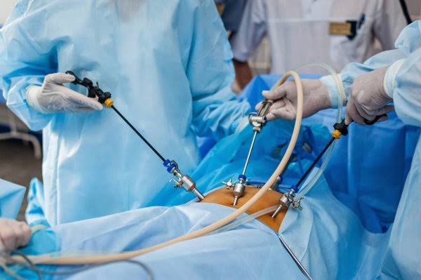
Similarly, a laparoscopy procedure is a keyhole to a woman’s abdomen and pelvic region and is especially useful to treat cysts, endometriosis and fibroids, in addition to a host of other conditions. This technique uses a laparoscope, a lightweight tool that is inserted through incisions made in the abdomen, to view an array of organs.
Laparoscopic treatment offers several advantages compared to traditional open surgery (laparotomy). These advantages include smaller incisions, reduced blood loss, shorter hospital stay, faster recovery, and potentially less post-operative pain.
Hysteroscopy
With the patient under anaesthesia, a thin viewing tool called a hysteroscope is inserted into the vagina and gently moved through the cervix into the uterus, where a liquid solution or carbon dioxide is inserted through the hysteroscope to expand the uterus. Once the uterus is expanded, a light and camera on the hysteroscope allow the doctor to see the endometrium (lining of the uterus), ovaries and fallopian tubes on a video screen.
Diagnostic hysteroscopy is used to identify problems in a woman’s uterus that may be contributing to infertility. The procedure may be performed to:
- Determine whether the uterus is abnormally shaped or contains scar tissue
- Find potential causes of recurrent miscarriages
- See blocked fallopian tubes
- Find fibroids and polyps
- Determine causes of abnormal bleeding and severe cramping.
Operative hysteroscopy is used to correct abnormal conditions that are determined during diagnostic hysteroscopy. Hysteroscopes may be used to insert small tools into the uterus to:
- Remove uterine fibroids and polyps
- Unblock fallopian tubes
- Remove the endometrium using a heated tool if endometriosis is present. Endometrial ablation is not performed in women who wish to become pregnant, as it destroys much of the lining of the uterus.
Laparoscopy
Laparoscopy for infertility is a minimally invasive surgical procedure that uses a laparoscope (a fibre-optic tube with light and video camera) inserted through two or more minor incisions, often in the belly button. The surgeon can then visually examine the pelvic reproductive organs and the pelvic cavity.
The procedure may be performed under general anaesthesia or local anaesthetic and typically takes 30 to 45 minutes. The abdomen is inflated with gas (carbon dioxide or nitrous oxide injected with a needle) to move the organs away from the abdominal wall so that they are visible during the procedure.
Once the abdomen is inflated, the laparoscope is inserted through the small incisions. The surgeon views the interior of the pelvic cavity on a video screen transmitting the images from the camera.
The surgeon will look for possible causes of infertility. These could be:
- Abnormalities of the uterus and ovaries
- Blocked fallopian tubes
- Scar tissue
- Fibroid tumours
- Endometriosis (which can only be confirmed via laparoscopy).
Common Symptoms
- Heavy or Prolonged Menstrual Bleeding: Heavy menstrual bleeding (menorrhagia) that may include prolonged periods or passing blood This can lead to anemia or fatigue.
- Pelvic Pain or Pressure: Pelvic pain or pressure ranging from mild discomfort to severe The pain may be constant or occur during menstruation or sexual intercourse.
- Enlarged Abdomen or Swelling: The abdomen to appear swollen or enlarged, similar to pregnancy.
- Frequent Urination: Fibroids or cysts that press on the bladder can result in increased frequency or urgency to urinate.
- Difficulty in Emptying the Bladder or Bowels: It can obstruct the bladder or bowels, causing difficulty in emptying them completely.
- Back or Leg Pain: It presses on nerves in the pelvis which may cause back pain or pain in the
- Infertility or Pregnancy Complications: Fibroids and ovarian cysts can interfere with the ability to conceive or cause complications during pregnancy, such as an increased risk of miscarriage, preterm labor, or breech presentation.
If you experience any symptoms that may be associated with uterine problems, it is advisable to consult with a healthcare provider or gynecologist. They can evaluate your symptoms, perform necessary tests, and recommend appropriate treatment options based on your individual circumstances.
How is it diagnosed?
Uterine factors are typically diagnosed through a combination of medical history, physical examination, and imaging tests. The process of diagnosing fibroids may include the following steps:
- Medical History: Your healthcare provider will ask you about your symptoms, menstrual history, and any family history of fibroids or related conditions.
- Pelvic Examination: A pelvic examination allows the healthcare provider to feel the size, shape, and position of your Fibroids can sometimes be felt as lumps or irregularities during this examination.
- Imaging Tests: Various imaging techniques can help visualize the presence, size, and location of fibroids or cysts. These tests may include:
- Ultrasound: Transabdominal or transvaginal ultrasound is commonly used to detect It uses sound waves to create images of the uterus and fibroids.
- MRI (Magnetic Resonance Imaging): MRI provides detailed images of the uterus, allowing for accurate identification of fibroids and their characteristics.
- Hysterosonography: This involves filling the uterus with saline and performing an ultrasound to get a clearer view within the uterine cavity.
- Other Tests: In some cases, additional tests may be recommended to rule out other conditions or gather more information. These may include hysteroscopy (a procedure using a thin tube with a camera to visualize the inside of the uterus), hysterosalpingography (an X- ray procedure to evaluate the shape and condition of the uterus and fallopian tubes), or blood tests to check for anemia or hormonal imbalances.
Based on the findings from these diagnostic procedures, your healthcare provider will determine if you have fibroids, their size, location, and characteristics. This information will guide treatment decisions and help develop an appropriate management plan tailored to your needs.
Laparoscopy for infertility is generally only performed after other fertility tests have not resulted in a conclusive diagnosis. For this reason, laparoscopy is often performed on women with unexplained infertility.
Laparoscopy also allows for biopsy of suspect growths and cysts that may be hampering fertility. Laparoscopy may be recommended for women experiencing pelvic pain, which is a potential symptom of endometriosis. Laparoscopy can also remove scar tissue that can be a cause of pelvic or abdominal pain.
What happens during a laparoscopic surgery?
Laparoscopic excision or laparoscopic surgery, is a minimally invasive surgical procedure used to diagnose and treat endometriosis. It involves making small incisions in the abdomen through which a laparoscope (a thin, lighted camera) and surgical instruments are inserted to visualize and remove endometrial implants and scar tissue.
Here’s an overview of the laparoscopic treatment of endometriosis:
- Anaesthesia: Before the procedure, anaesthesia is administered to ensure the patient’s comfort and pain General anaesthesia is typically used, which means you will be asleep during the surgery.
- Creation of Incisions: Several small incisions, usually around 5 to 1 centimetre in length, are made in the abdomen. These serve as entry points for the laparoscope and surgical instruments.
- Insertion of Laparoscope: The laparoscope, a thin tube with a light and camera at the end, is inserted through one of the incisions. It allows the surgeon to visualize the pelvic organs, including the uterus, ovaries, fallopian tubes, and surrounding structures, on a monitor.
- Exploration and Evaluation: The surgeon carefully examines the pelvic area to identify any endometrial implants, adhesions, or cysts associated with endometriosis. The extent and severity of the endometriosis are assessed.
- Excision and Removal: Using specialized surgical instruments, the surgeon removes or excises the endometrial implants, scar tissue, and any other abnormalities found during the exploration. The goal is to remove as much endometriosis tissue as possible while preserving healthy tissue.
- Closure and Recovery: Once the necessary excisions are complete, the incisions are closed with sutures or surgical tape. Some surgeons may use dissolvable stitches that do not require removal. The patient is then moved to a recovery area, where they are closely monitored until the effects of anaesthesia wear off.
What is the recovery like?
The recovery period after surgery can vary depending on the extent of the procedure performed, individual factors, and the overall health of the patient. Here are some general aspects of the recovery process:
- Hospital Stay: The length of the hospital stay will depend on the type of procedure performed. If it was a laparoscopic procedure, which is minimally invasive, the hospital stay is typically short, often just a few hours or overnight. In more complex cases or if open surgery (laparotomy) was required, a longer hospital stay may be necessary, usually a few
- Pain Management: Pain and discomfort are common after endometriosis surgery. Your healthcare provider will prescribe pain medication to help manage post-operative It’s important to take the medication as directed and report any severe or persistent pain to your healthcare team.
- Activity and Rest: It’s essential to allow your body to heal and recover after surgery. Your healthcare provider will provide specific instructions on when and how to resume activities, including work, exercise, and lifting heavy objects. Initially, you may need to limit physical activity and take time off from work to rest and recover. Gradually, you can increase your activity level as advised by your healthcare provider.
- Wound Care: If you had laparoscopic surgery, you will have small incisions that require proper care. Keep the incision sites clean and dry as instructed by your healthcare provider. They will provide guidance on when to remove any dressings and when it’s safe to shower or
- Follow-Up Appointments: You will have follow-up appointments scheduled with your healthcare provider to monitor your recovery progress. These appointments allow your healthcare team to assess your healing, address any concerns or complications, and discuss long-term management options if needed.
- Emotional Support: Coping with a chronic condition like endometriosis and going through surgery can be emotionally It’s important to seek emotional support from loved ones, support groups, or mental health professionals who can provide guidance, understanding, and coping strategies.
Recovery time varies for each individual, but most people can expect to resume their regular activities within a few weeks to a couple of months after surgery, depending on the extent of the procedure. It’s crucial to follow your healthcare provider’s instructions, take care of yourself, and communicate any concerns or unexpected symptoms during the recovery period.
Remember that the recovery experience can be different for everyone, and it’s important to consult with your healthcare provider for personalized advice and guidance based on your specific situation.

